Comparative Analysis of Pre-Trained Deep Learning Models for Classifying Tropical Fungal Skin Infections
DOI:
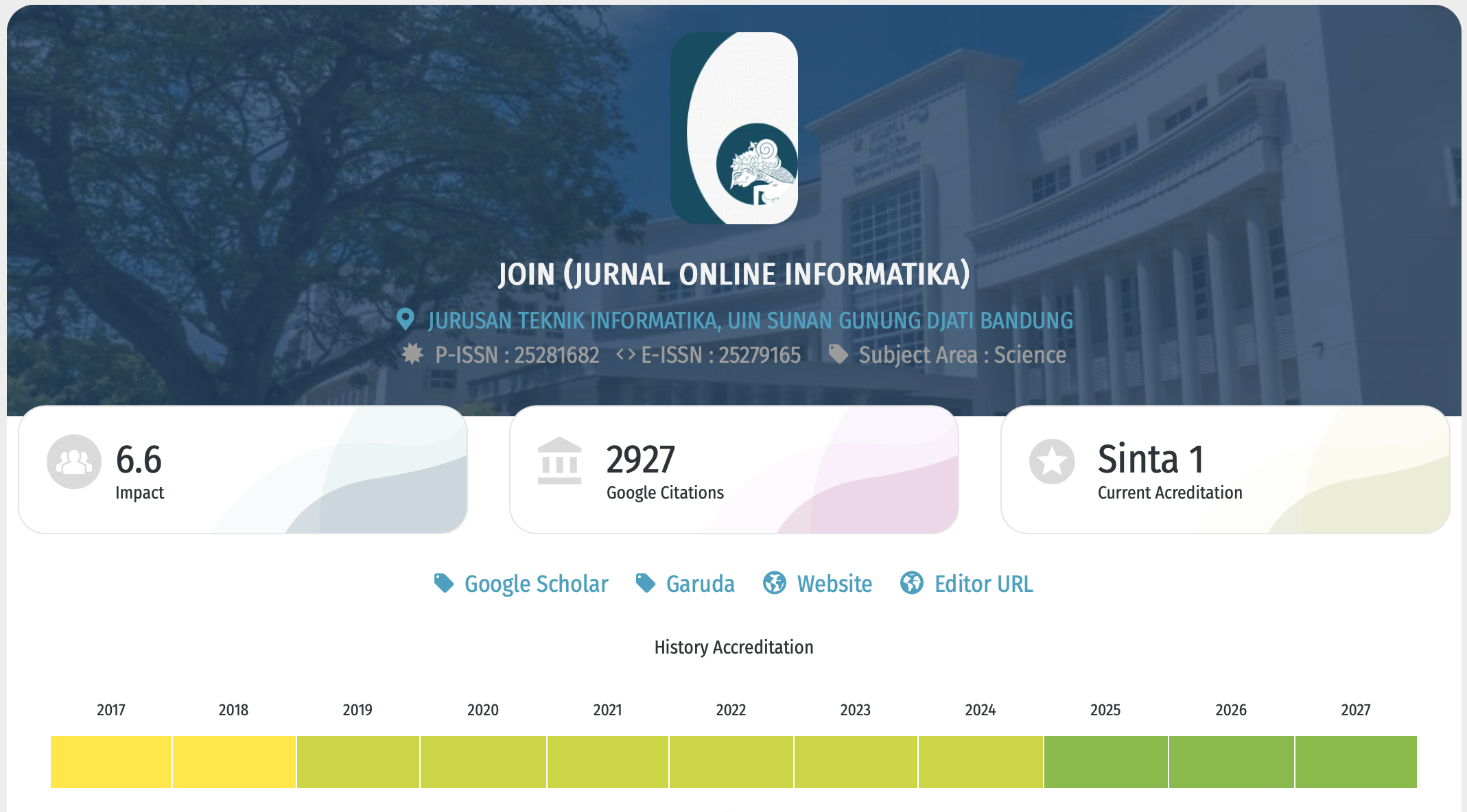
https://doi.org/10.15575/join.v11i1.1758Keywords:
Deep Learning, Fungal Skin Infections, Medical Image Classification, MobileNetV3, Tropical Skin InfectionsAbstract
References
[1] P. N. Srinivasu, J. G. Sivasai, M. F. Ijaz, A. K. Bhoi, W. Kim, and J. J. Kang, “Classification of skin disease using deep learning neural networks with mobilenet v2 and lstm,” Sensors, vol. 21, no. 8, Apr. 2021, doi: 10.3390/s21082852.
[2] D. Li et al., “Worldwide trends and future projections of fungal skin disease burden: a comprehensive analysis from the Global Burden of Diseases study 2021,” Front. Public Health, vol. 13, 2025, doi: 10.3389/fpubh.2025.1580221.
[3] World Health Organization (WHO), “Report of the first WHO global meeting on skin-related neglected tropical diseases,” World Health Organization (WHO), 2023. Accessed: Oct. 06, 2025. [Online]. Available: https://www.who.int/publications/i/item/9789240081201
[4] Ministry of Health and Republic of Indonesia (Kemenkes RI), “Laporan Nasional Riskesdas,” Ministry of Health, Republic of Indonesia (Kemenkes RI), Jakarta, 2019. Accessed: Jul. 06, 2025. [Online]. Available: https://www.litbang.kemkes.go.id/laporan-riskesdas-2019/
[5] S. Azzahra, E. Ervianti, and R. Setiabudi, “Clinical Patterns and Demographic Characteristics of Dermatophytosis in Surabaya,” Indonesian Journal of Tropical and Infectious Disease, vol. 12, no. 3, pp. 252–267, Dec. 2024, doi: 10.20473/ijtid.v12i3.66511.
[6] M. F. Petrucelli et al., “Epidemiology and diagnostic perspectives of dermatophytoses,” Journal of Fungi, vol. 6, no. 4, pp. 1–15, Dec. 2020, doi: 10.3390/jof6040310.
[7] V. Ravi, “Attention Cost-Sensitive Deep Learning-Based Approach for Skin Cancer Detection and Classification,” Cancers (Basel)., vol. 14, no. 23, Dec. 2022, doi: 10.3390/cancers14235872.
[8] M. K. Khalifaturohman, “Systematic Literature Review: Deep Learning Models in Arabic Script Classification,” Khazanah Journal of Religion and Technology, vol. 3, no. 1, pp. 18–22, Jul. 2025, doi: 10.15575/kjrt.v3i1.1619.
[9] M. Alruwaili and M. Mohamed, “AEfficientNetn Integrated Deep Learning Model with and ResNet for Accurate Multi-Class Skin Disease Classification,” Diagnostics, vol. 15, no. 5, Mar. 2025, doi: 10.3390/diagnostics15050551.
[10] A. Nur, A. Thohari, L. Triyono, I. Hestiningsih, B. Suyanto, and A. Yobioktobera, “Performance Evaluation of Pre-Trained Convolutional Neural Network Model for Skin Disease Classification,” JUITA: Jurnal Informatika, vol. 10, no. 1, pp. 9–18, 2022, Accessed: Oct. 06, 2025. [Online]. Available: https://jurnal.stmiktime.ac.id/index.php/juita/article/view/802
[11] T. Alyas, K. Alissa, A. S. Mohammad, S. Asif, T. Faiz, and G. Ahmed, “Innovative Fungal Disease Diagnosis System Using Convolutional Neural Network,” Computers, Materials and Continua, vol. 73, no. 3, pp. 4869–4883, 2022, doi: 10.32604/cmc.2022.031376.
[12] T. D. Nigat, T. M. Sitote, and B. M. Gedefaw, “Fungal Skin Disease Classification Using the Convolutional Neural Network,” J. Healthc. Eng., vol. 2023, 2023, doi: 10.1155/2023/6370416.
[13] T. Debebe and B. Molla, “Human Skin Fungal Diseases Classification Using Deep Learning Technique,” Haramaya Journal of Engineering and Technology (HJET), vol. 1, no. 2, pp. 16–37, 2022, [Online]. Available: https://journals.ddu.edu.et/index.php/HJET
[14] E. da S. Puls, M. V. Todescato, and J. L. Carbonera, “An evaluation of pre-trained models for feature extraction in image classification,” Oct. 2023, [Online]. Available: http://arxiv.org/abs/2310.02037
[15] P. Chanyachailert, C. Leeyaphan, and S. Bunyaratavej, “Cutaneous Fungal Infections Caused by Dermatophytes and Non-Dermatophytes: An Updated Comprehensive Review of Epidemiology, Clinical Presentations, and Diagnostic Testing,” Journal of Fungi, vol. 9, no. 6, Jun. 2023, doi: 10.3390/jof9060669.
[16] N. Łabędź, C. Navarrete-Dechent, H. Kubisiak-Rzepczyk, M. Bowszyc-Dmochowska, A. Pogorzelska-Antkowiak, and P. Pietkiewicz, “Pityriasis Versicolor—A Narrative Review on the Diagnosis and Management,” Oct. 01, 2023, Multidisciplinary Digital Publishing Institute (MDPI). doi: 10.3390/life13102097.
[17] Q. Yu et al., “Clinical Analysis of 57 Patients With Interdigital Infection in Shanghai, China: A Cross-Sectional Study,” Int. J. Dermatol. Venereol., vol. 8, no. 2, pp. 100–105, Jun. 2025, doi: 10.1097/JD9.0000000000000233.
[18] A. K. C. Leung, J. M. Lam, K. F. Leong, and K. L. Hon, “Tinea corporis: An updated review,” Jul. 20, 2020, Bioexcel Publishing LTD. doi: 10.7573/dic.2020-5-6.
[19] M. Trigka and E. Dritsas, “A Comprehensive Survey of Deep Learning Approaches in Image Processing,” Sensors (Basel), vol. 25, no. 2, Jan. 2025, doi: 10.3390/s25020531.
[20] D. Murcia-Gómez, I. Rojas-Valenzuela, and O. Valenzuela, “Impact of Image Preprocessing Methods and Deep Learning Models for Classifying Histopathological Breast Cancer Images,” Applied Sciences (Switzerland), vol. 12, no. 22, Nov. 2022, doi: 10.3390/app122211375.
[21] M. Tan and Q. V. Le, “EfficientNet: Rethinking Model Scaling for Convolutional Neural Networks,” arXiv preprint arXiv:1905.11946, no. 6, Sep. 2020, doi: 10.48550/arXiv.1905.11946.
[22] A. Howard et al., “Searching for MobileNetV3,” in Proceedings of the IEEE/CVF International Conference on Computer Vision (ICCV), IEEE, 2019. doi: 10.48550/arXiv.1905.02244.
[23] I. Manole, A. I. Butacu, R. N. Bejan, and G. S. Tiplica, “Enhancing Dermatological Diagnostics with EfficientNet: A Deep Learning Approach,” Bioengineering, vol. 11, no. 8, Aug. 2024, doi: 10.3390/bioengineering11080810.
[24] S. Thirumalaisamy et al., “Breast Cancer Classification Using Synthesized Deep Learning Model with Metaheuristic Optimization Algorithm,” Diagnostics, vol. 13, no. 18, Sep. 2023, doi: 10.3390/diagnostics13182925.
[25] B. Ashwini, M. Kaur, D. Singh, S. Roy, and M. Amoon, “Efficient Skip Connections-Based Residual Network (ESRNet) for Brain Tumor Classification,” Diagnostics, vol. 13, no. 20, Oct. 2023, doi: 10.3390/diagnostics13203234.
[26] M. Huang and Y. Xu, “Image classification of Chinese medicinal flowers based on convolutional neural network,” Mathematical Biosciences and Engineering, vol. 20, no. 8, pp. 14978–14994, 2023, doi: 10.3934/mbe.2023671.
[27] S. zhenlin, “Lightweight Face Anti-spoofing for Improved MobileNetV3,” Journal of Image Processing Theory and Applications, vol. 7, no. 1, 2024, doi: 10.23977/jipta.2024.070117.
[28] J. Mohan, A. Sivasubramanian, V. Sowmya, and R. Vinayakumar, “Enhancing Skin Disease Classification Leveraging Transformer-based Deep Learning Architectures and Explainable AI,” Comput. Biol. Med., Jul. 2024, doi: 10.1016/j.compbiomed.2025.110007.
[29] M. A. Khan, M. Sharif, T. Akram, R. Damaševičius, and R. Maskeliūnas, “Skin lesion segmentation and multiclass classification using deep learning features and improved moth flame optimization,” Diagnostics, vol. 11, no. 5, 2021, doi: 10.3390/diagnostics11050811.
Downloads
Published
Issue
Section
Citation Check
License
Copyright (c) 2026 Suhardi Aras, Muhammad RIzwan Darwis, Muhammad Zhaky Arkan

This work is licensed under a Creative Commons Attribution-NoDerivatives 4.0 International License.
You are free to:
- Share — copy and redistribute the material in any medium or format for any purpose, even commercially.
- The licensor cannot revoke these freedoms as long as you follow the license terms.
Under the following terms:
-
Attribution — You must give appropriate credit, provide a link to the license, and indicate if changes were made. You may do so in any reasonable manner, but not in any way that suggests the licensor endorses you or your use.
-
NoDerivatives — If you remix, transform, or build upon the material, you may not distribute the modified material.
-
No additional restrictions — You may not apply legal terms or technological measures that legally restrict others from doing anything the license permits.
Notices:
- You do not have to comply with the license for elements of the material in the public domain or where your use is permitted by an applicable exception or limitation.
- No warranties are given. The license may not give you all of the permissions necessary for your intended use. For example, other rights such as publicity, privacy, or moral rights may limit how you use the material.

This work is licensed under a Creative Commons Attribution-NoDerivatives 4.0 International License